03/27/2019 Alena Masheva Health
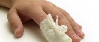
A woman's hands are her calling card. They are always in sight. Therefore, for most beauties, a weekly manicure in the salon is a mandatory procedure. Unfortunately, clients rarely think about the fact that their next visit to a hairdresser could end badly. The procedure can cause the development of a pathology such as subcutaneous panaritium. The disease often leads to the removal of the nail. It also provokes the development of even more serious complications.
Purulent inflammation of the finger
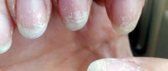
At the very base of the nail plate there is a small area of living skin. This is an eponychium. But most people call it cuticle. This epithelial thin film surrounds the nail plate in the area of the hole. Its main role is protective. The thin skin is a barrier to foreign bodies and bacteria. Thanks to the cuticle, they are not able to penetrate into the growth zone of the nail.
Trimming the eponychium is not recommended. If you do this, it begins to grow stronger and becomes rougher. A similar mechanism can be observed in the formation of scars.
If for some reason the eponychium is damaged, the gates to infection open. Most often this happens during a manicure. In addition, it can be observed during games in children. Pyogenic microbes penetrate into the tissues. For example: staphylococcus or streptococcus. Sometimes mixed microflora is also diagnosed.
In most cases, the localization of subcutaneous panaritium occurs on the palmar surface. However, the swelling is more noticeable on the back of the finger.
Under the skin of the palms there are very dense tendon cords. They intertwine with bundles of connective tissue and create cells. In their structure they resemble a honeycomb. Each cell is filled with fat. This structure contributes to the fact that the inflammatory process does not spread along the plane, but goes deeper. This poses a risk to tendons, joints and bones.
The inflammatory effusion, due to the conditions listed above, is under severe pressure. This provokes the appearance of acute, throbbing pain. The accumulation of exudate impairs blood circulation. Compression of the blood vessels occurs, which can lead to tissue necrosis.
Operation
The operation of felon on the finger in our clinic is carried out on an outpatient basis, by experienced surgeons using modern techniques and high-tech equipment. There are two main methods of treating felon in the form of surgery:
- surgical removal;
- Laser removal of felon.
Both methods involve opening a purulent formation under the skin or nail plate and completely removing the pus. After this, the wound is sanitized with an antiseptic (if a laser is used, the edges of the wound are cauterized).
If the nail plate comes off, then partial or complete removal of the fingernail is performed during panaritium.
Factors that increase the risk of developing felon
Subcutaneous panaritium of the finger can develop only if infection penetrates into the soft tissue. Most often, staphylococcus is the culprit of the pathology. In addition, intestinal, gram-positive and gram-negative bacilli can cause the development of the disease; anaerobic non-clostridial microflora; Proteus; putrefactive infection and streptococcus.
Doctors say that subcutaneous panaritium of a finger on the hand is most often observed in children. And also in people aged 20 to 50 years. According to statistics, 30% of patients get sick due to minor injuries they receive at work. In most cases, the infection is localized on the index, middle and ring fingers of the right hand.
The following factors contribute to the development of infection:
- Diabetes.
- Habit of biting your nails or biting your fingertips.
- Washing hands using chemicals or some types of soap.
- Immunodeficiency states.
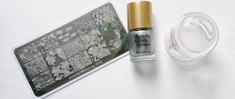
- Abuse of cosmetic procedures that can injure the nail or skin of the finger. For example: manicure or pedicure.
- Avitaminosis. Subcutaneous panaritium of the finger often develops in people who are forced to take medications with vitamin A or its derivatives. Such medications can negatively affect the immune system.
- Chemotherapy.
- Lupus erythematosus, psoriasis. As well as other chronic skin diseases.
- Taking immunosuppressants.
- Hypothermia.
- Frequent exposure to vibration.
- Vascular diseases of the extremities.
- Introduction of a foreign body. For example, a small pebble or sliver.
- Mycosis of the foot or nail.
- Hyperhidrosis.
- Burn.
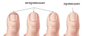
- Ingrown nail.
Causes of panaritium
In addition to skin contamination, some internal factors of the body contribute to the development of felon:
- Exposure of the skin to various irritants, chemicals and metals.
- Hypothermia.
- Often, an increased tendency to develop panaritium occurs in some common diseases: diabetes mellitus, vitamin deficiency, changes in metabolism and the functioning of the immune system.
In these conditions, tissue nutrition and blood supply are disrupted. Therefore, it is easier for a pathogen to penetrate through microtraumas on the skin of the fingers and toes.
The most common cause of panaritium is staphylococcus. Less commonly, streptococci, Proteus, Pseudomonas aeruginosa and other microorganisms lead to its development.
The infection penetrates through small puncture wounds on the palmar surface of the skin of the finger on fish bones, metal shavings, and wood chips. Or through abrasions, skin cracks, minor burns, manicure wounds and other minor wounds.
Since the wounds are small, patients often do not pay attention to them and do not treat them in a timely manner. And, taking into account the structural features of the skin and blood supply to the hand, a small wound channel closes very quickly. Therefore, the infection remains in the wound, leading to the formation of inflammatory fluid (pus).
The liquid, unable to flow out of the wound, rushes deeper along the bridges of the subcutaneous fat layer. It involves muscles, ligaments, tendons and their sheaths, joints, and bones in the inflammatory process. Depending on the place of origin and the route of spread of the inflammatory process, several types of panaritium are distinguished.
Classification
Subcutaneous panaritium (ICD code 10 L03.0) was classified by doctors as a particular form of cellulite. Depending on the location of the inflammation and the nature of the lesion, several types of pathology are distinguished:
- Skin. This is the lightest, most superficial form. The abscess is localized in the thickness of the skin. In appearance it resembles a blister. Sometimes you can see a cavity with yellow pus and blood. There is hyperemia around the lesion.
- Subcutaneous panaritium. This is the most common form. As a rule, inflammation is concentrated on the nail phalanx. Over time, it can spread to others. The finger is swollen. Necrosis and purulent melting of the fiber may be observed. Finger mobility is limited. The pain is sharp and throbbing. Possible increase in body temperature. In the event that conservative treatment does not produce results, an opening of the subcutaneous panaritium is required. It must be urgently carried out after the patient's first sleepless night. Palpation helps to correctly determine the point of greatest pathological changes in tissue.
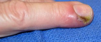
- Paronychia. Inflammation of the nail fold. This disease can be caused by hangnails. The cushion becomes painful, turns red and swells.
- Subungual panaritium. The cause of development may be a splinter, a puncture wound or a hematoma. It is often the result of the development of paronychia. The roller swells. When you press on it, pus may be released.
- Bone. There is a primary type, which develops as a result of puncture wounds. Provided that the periosteum is damaged. Secondary occurs as a complication of subcutaneous. With this pathology, the patient’s body temperature can rise to 40 degrees. Chills and severe headache are noted. Destructive changes in the phalanges can be detected on an x-ray on the 10th day of illness. If conservative treatment does not produce results within 48 hours, surgical intervention is required.
- Articular. Fusiform swelling, hyperemia and pain in the finger develop. The infection first affects soft tissue. Gradually it affects the cartilage and surfaces of the interphalangeal joints. Destruction of ligaments can provoke the appearance of crunching and pathological mobility.
- Tendinous. It is considered the most dangerous form. The cause of the development of this pathology may be subcutaneous panaritium. As well as infection through a puncture. Purulent inflammation affects the tendons and their connective membranes. Immediate intervention by an experienced surgeon is required.
All of these types are diagnosed both individually and in combination. Most often, patients are affected by the subcutaneous form. If the patient ignores the problem, the pathology begins to progress. In this case, it poses a real threat to health and even life.
Types of felon
Cutaneous panaritium occurs on the back of the finger (near the nail). Pus accumulates under the skin and forms a blister. The skin around it may become very red. The pain in this type of disease is very mild, sometimes a burning sensation may occur. After some time, the bubble enlarges, which means that the disease progresses and spreads deeper into the tissue.
Periungual panaritium occurs on the periungual ridge. First, inflammation appears at the edge of the nail plate. If the inflammation is not treated, it will spread further.
Subungual panaritium is slightly less common. In this case, pus accumulates under the nail plate. This may be the result of pus spreading across the finger or simply a sore where the nail meets the finger.
When inflammation begins on the surface of the finger from the palmar side, it is called subcutaneous panaritium. On this side the skin is much denser and pus cannot come to the surface. Because of this, the inflammation process goes deep and can reach the bone.
Bone panaritium is expressed as inflammation of the bone. This may be due to an open fracture, when the infection enters directly into the bone, or it may be a consequence of advanced panaritium of previous forms.
Articular panaritium most often affects the joint of the first phalanx. It can also occur due to injury or prolonged purulent processes in the tissues of the finger. The patient feels pain in the finger upon palpation or movement.
The consequence of articular felon can be osteoarticular felon. It affects the articular ends of the phalanges of the fingers. In this case, the tendons remain intact.
But with tendon panaritium (or tenosynovitis), the tendons are already affected. This leads to impaired hand function. In this case, the finger is in a bent position and the patient feels severe pain. Bone, articular, osteoarticular and tendon forms of the disease are considered deep (severe) and require surgical intervention.
There are several types of felon
Panaritium occurs not only on the fingers, but also on the toes. The panaritium of the toe essentially looks the same as on the hands. Fortunately, the disease can be easily detected. When panaritium occurs on the leg, swelling and inflammation occur near the nail, and the patient feels pain.
The likelihood of developing panaritium in children is even higher than in adults. After all, children do not care so much about hygiene. And the smallest ones can get panaritium due to carelessly cut nails. In addition, in children the protective functions of the body are less developed than in adults and it is easier for various infections to develop.
Stages
The subcutaneous panaritium of the finger on the hand has three stages of development. Only a doctor can accurately determine it. The treatment tactics he chooses depends on this. The first stage is often asymptomatic. The infection penetrates the soft tissue and begins to multiply. The only thing the patient can feel is itching at the site of penetration of the bacterial flora.
In the second stage, cellular elements mixed with lymph and blood begin to accumulate in the affected tissue. The inflammatory infiltrate contains lymphocytes, histiocytes, and erythrocytes. As well as lymphoid and plasma cells. The tissues swell. The patient feels severe pain.
In the third stage, abscess formation occurs. Melting of the inflamed tissues is observed. A cavity is formed in which pus accumulates.
Conservative therapy is effective in the first and second stages. Under the supervision of a doctor, treatment can be carried out at home. But if an abscess has formed, surgical intervention will be required. In the third stage, conservative treatment is no longer effective.
Symptoms
The first stage of felon is usually asymptomatic. Subsequently, signs of pathology begin to gradually appear and intensify. The main symptoms of this disease include:
- Hyperemia and edema.
- Malaise.
- Low-grade fever.
- Feeling of fullness in the finger.
- Pain on palpation at the site of inflammation. Most often, the discomfort intensifies at night. Throbbing pain almost always accompanies subcutaneous panaritium. Treatment in this case is required immediately.
- Local increase in temperature.
- Decreased motor function of the phalanx.
- Enlarged lymph nodes.
- Headache.
- In severe cases, severe intoxication is observed. The patient may suffer from dizziness and nausea.
Diagnostics
Sharp throbbing pain, redness and swelling in superficial forms of panaritium allow you to immediately make a diagnosis and begin treatment. When deep tissues - bones, joints and tendons - are involved in the pathological process, diagnosis is made difficult by the blurred clinical picture and the absence of characteristic symptoms. To clarify the diagnosis and obtain reliable data on the location of the disease, radiography is used.
Intense pain and swelling that appeared at the very beginning of the disease are not clearly reflected on x-rays - the soulless device registers only obvious changes in the hard core of the finger. Therefore, the procedure often has to be repeated at intervals of 1-2 weeks.
Possible complications
Treatment of subcutaneous panaritium of the finger should not be delayed. It should be carried out immediately under the supervision of a doctor. If the patient ignores the first symptoms of the pathology and delays treatment, the disease progresses quickly. Conservative therapy is no longer able to help in the third stage.
Panaritium is not a harmless disease, as many people think. It can provoke the development of the following complications:
- Inflammation of lymphatic vessels and nodes.
- Sepsis.
- Inflammatory muscle damage.
- Vascular thrombosis. As well as inflammation of the walls of the veins.
- Osteomyelitis.
- Gangrene of the finger.
The pus can spread to the hand and even forearm. This condition poses a real threat to the patient’s life. In this case, amputation of the finger may be required.
Forecast
If medical assistance was provided in the initial stages of the disease, the prognosis for a full recovery is positive. If the inflammatory process has progressed to the second or third stage, even after successful excision of the suppuration, the motor function of the finger will be limited by stiffness. Ankyloses are less common.
In the initial stages, felon may seem like a harmless redness, like after an unsuccessful hangnail. Despite this, you should not put off visiting a doctor or self-medicate. Damage to the finger requires qualified treatment , and in some cases even surgical intervention.
Surgery
Purulent surgeons often encounter in their practice such a problem as subcutaneous panaritium. Surgery is prescribed if the disease has progressed to the third stage. Most often, the surgeon prescribes surgery immediately after the patient's first sleepless night due to severe pain.
The procedure is performed under general anesthesia or regional anesthesia. It should only be performed by an experienced doctor. Otherwise, it will not be possible to completely relieve the pain syndrome. This may negatively affect the success of the operation. Patients who have previously undergone a similar procedure remember the feeling of pain well. They wait with horror for a repetition of these sensations. Often they even refuse surgery. Therefore, the doctor’s first priority is to completely anesthetize the affected area.
After the anesthesia has begun to take effect, the doctor begins surgical treatment of subcutaneous panaritium. Incisions should be made directly above the center of the purulent-necrotic lesion. The cavity is washed. For this, Dimexide, Chlorhexidine or Furosimide are used. A drainage is installed to drain the pus. It is made from a small piece of medical gum.
Many modern surgeons refuse to use drainage. During surgery, they cut out a spindle-shaped strip of subcutaneous fat within the healthy tissue. Such an open, crater-shaped wound heals much faster and does not provoke the development of complications. The same cannot be said about side incisions that require drainage. Upon completion of the operation, a bandage with Levomekol or another drug chosen by the surgeon is applied.
During rehabilitation, the attending physician prescribes antibiotics. The dressing should be changed daily. In the first days, ointments are used to help draw out the pus. Subsequently, antibacterial agents are used.
During the rehabilitation period, physical procedures may be prescribed:
- Electrophoresis.
- UHF.
- Ural Federal District.
Pharmacy drugs
Conservative therapy will be effective in the first stages of pathology development if the patient consults a doctor in time. Most likely, surgery will not be needed. The doctor will confirm the diagnosis and give detailed instructions on how to treat subcutaneous panaritium with medications.
The following drugs have proven themselves best in the fight against pathology:
- Azithromycin. The duration of treatment is three days. Once a day, the patient should take a tablet containing 500 mg of the active substance. If necessary, the dose can be increased by the doctor.
- "Sumalek." The patient should take 0.5 g of the active substance once a day. The duration of treatment is determined by the doctor.
- "Ziromin." The drug allows you to cure felon in three to five days. The recommended dose is 500 mg once daily.
- Ichthyol ointment. This is a traditional remedy in the fight against panaritium. The drug promotes the release of pus. The ointment is used for dressings, which should be changed at least three times a day.
- "Dimexide". Before use, the drug is diluted with water in a ratio of 1:4. Gauze is soaked in liquid and used as a compress. The duration of the procedure is 40 minutes.
- "Levomekol". The ointment is used as a compress. The drug helps cleanse tissues of pus. In addition, it destroys pathogenic flora. "Levomekol" is used as a compress, which is applied twice a day.
- Vishnevsky ointment. This is one of the most effective remedies that can cure subcutaneous panaritium. Photos taken by patients confirm that swelling and hyperemia decrease within a few hours after the start of treatment. This occurs due to the rapid blocking of inflammation. The ointment helps open the abscess and cleanse the wound. It is used twice a day for compresses.
- Tetracycline ointment. This antibacterial agent relieves pain, hyperemia and inflammation well. The ointment is applied twice a day for ten days. A thin layer of it is applied to the site of inflammation. In order to enhance effectiveness, tetracycline ointment can be alternated with zinc ointment.
- "Dermasept" (gel). A sterile gauze pad is impregnated with the drug and applied to the site of inflammation. Dermasept gel can be used four times a day.
- Syntomycin ointment. The drug is used for a bandage that is applied at night. The amount of ointment for one procedure should not exceed the size of a pea. Treatment with this drug should not last longer than two weeks.
Traditional methods
Many traditional medicine recipes allow you to painlessly eliminate subcutaneous panaritium. Treatment at home should be carried out with the consent and supervision of a doctor. This will avoid the development of unwanted effects.
The most effective recipes include:
- Pass eight to ten cloves of garlic through a press. Pour the resulting slurry with a glass of hot water, the temperature of which is 80 degrees. The liquid should be infused for seven minutes. The affected finger is dipped into the solution for a few seconds. The manipulation is repeated until the liquid cools down. You can also use baths with crumbs of laundry soap, soda, copper sulfate, salt and celandine. The procedure should be repeated three times a day.
- The juice or pulp of aloe leaves is used for compresses.
- Castor oil is heated in a water bath to a temperature of 40 degrees. A gauze pad is soaked in a warm solution and applied to the site of inflammation. Cover with cellophane and insulate. The compress is left for two hours.
- A quarter of an onion head and four cloves of garlic are passed through a meat grinder. The resulting pulp is used for compresses.
- One hundred grams of raw beets are ground on a fine grater. Add 50 g of fat sour cream. The resulting mixture is applied to the affected area and covered with a bandage.
Medicines for the treatment of panaritium
Medications are necessary at all stages of development of subungual felon, as it is provoked by pyogenic microbes. It is simply impossible to suppress them without chemicals and antiseptics.
Baths
These procedures are carried out provided that the inflammatory process is just beginning and the pain is still mild.
An effective alternation method:
- With potassium permanganate (potassium permanganate), which acts as a strong antiseptic. The solution is made weak. The water temperature is warm. The finger is kept in the solution for up to half an hour.
- With calendula, use a ready-made tincture in alcohol. It has anti-inflammatory properties. Dilute in warm water (100ml:10 ml). The procedure time is similar.
- With eucalyptus (alcohol tincture). Suppresses the growth of Staphylococcus aureus. Dilute in warm water (100ml:10ml). The procedure takes up to half an hour.
After the last bath, a gauze cloth is moistened with eucalyptus tincture and applied to the site of inflammation.
Treatment with antibiotics
When infected with purulent microbes, antibiotics from the penicillin group are prescribed.
This usually occurs in acute form of panaritium, when the patient is in the hospital and receives surgical treatment.
For fungal infections, antifungal medications are recommended.
If the disease is mild, then antibiotics for oral, intravenous, and intramuscular use are not prescribed. They can only be recommended topically, in the form of a soft dosage form.
Ointment
Tetracycline, Levomikol, Lincomycin are antibiotic ointments that act on purulent microbes. After a warm bath, which increases blood flow to the lesion, the ointment is applied in a thick layer in the form of an application. Secure tightly with a bandage.
Prevention
Most people begin to value their health only after they get sick. It is difficult for women to imagine that such an ordinary procedure as a manicure can cause the development of gangrene and loss of a finger. Fortunately, such cases are quite rare. However, you need to take maintaining your health seriously.
Prevention is always preferable to treatment. Subcutaneous panaritium of a finger on the hand is one of those pathologies that are quite easy to prevent. To do this, just adhere to the following rules:
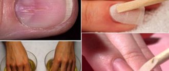
- Always wash your hands thoroughly with soap.
- Treat any skin damage, even the most minor, with peroxide or chlorhexidine. Cover wounds with a bactericidal plaster.
- Use individual manicure tools or visit only a trusted specialist.
- When working with soil, always wear textile-lined rubber gloves.
- Monitor the condition of the cuticle and prevent the appearance of hangnails.
Source: fb.ru